Temiz, Hakan
Loading...
Profile URL
Name Variants
Temiz, Hakan, Temiz, H., Hakan Temiz
Job Title
Prof. Dr.
Email Address
hakantemiz@artuklu.edutr
Main Affiliation
Department of Basic Medical Sciences / Temel Tıp Bilimleri Bölümü
Status
ORCID ID
Scopus Author ID
Turkish CoHE Profile ID
Google Scholar ID
WoS Researcher ID
Sustainable Development Goals
 1
1NO POVERTY
0
Research Products
 2
2ZERO HUNGER
0
Research Products
 3
3GOOD HEALTH AND WELL-BEING
2
Research Products
 4
4QUALITY EDUCATION
0
Research Products
 5
5GENDER EQUALITY
0
Research Products
 6
6CLEAN WATER AND SANITATION
0
Research Products
 7
7AFFORDABLE AND CLEAN ENERGY
0
Research Products
 8
8DECENT WORK AND ECONOMIC GROWTH
0
Research Products
 9
9INDUSTRY, INNOVATION AND INFRASTRUCTURE
0
Research Products
 10
10REDUCED INEQUALITIES
0
Research Products
 11
11SUSTAINABLE CITIES AND COMMUNITIES
0
Research Products
 12
12RESPONSIBLE CONSUMPTION AND PRODUCTION
0
Research Products
 13
13CLIMATE ACTION
0
Research Products
 14
14LIFE BELOW WATER
1
Research Products
 15
15LIFE ON LAND
0
Research Products
 16
16PEACE, JUSTICE AND STRONG INSTITUTIONS
0
Research Products
 17
17PARTNERSHIPS FOR THE GOALS
0
Research Products
Documents
18
Citations
70
h-index
5
Documents
13
Citations
71
Scholarly Output
3
Articles
3
Views / Downloads
2/40
Supervised MSc Theses
0
Supervised PhD Theses
0
WoS Citation Count
6
Scopus Citation Count
5
Patents
0
Projects
0
WoS Citations per Publication
2.00
Scopus Citations per Publication
1.67
Open Access Source
3
Supervised Theses
0
| Journal | Count |
|---|---|
| European Review for Medical and Pharmacological Sciences | 1 |
| Northwestern Medical Journal | 1 |
| Progress in Nutrition | 1 |
Current Page: 1 / 1
Scopus Quartile Distribution
Competency Cloud
3 results
Scholarly Output Search Results
Now showing 1 - 3 of 3
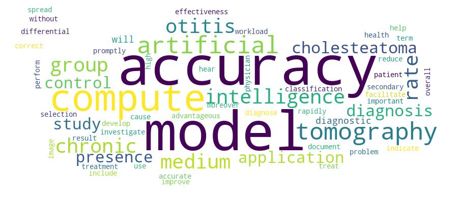
Article An Investigation of the Role of Trace Elements and Biochemical Parameters in Patients With COVID-19(2025) Erkan, Revşa Evin Canpolat; Unsal, Velıd; Özbek, Erdal; Sabancılar, İlhan; Temız, Hakan; Mermutluoğlu, ÇiğdemAim: The COVID-19 pandemic is an emergent viral respiratory disease characterized by high fever and shortness of breath, and it was declared a pandemic by the World Health Organization in March 2020. Early assessment of patients’ biochemical tests is important for accelerating diagnosis, allowing effective treatment, and controlling the further spread of the disease. The present study aimed to investigate the association between the disease, trace elements -including copper (Cu), zinc (Zn), selenium (Se), manganese (Mn), and cobalt (Co) vitamin D, Alanine aminotransferase (ALT) and Aspartate aminotransferase (AST) biochemical levels, and the correlation between the parameters tested in patients with COVID-19. Methods: In our study, 40 patients (case group) who were hospitalized with a diagnosis of COVID-19 based on chest X-ray images and RT-PCR results evaluated by an infectious diseases specialist were included, along with 40 healthy individuals (control group) over the age of 18 who had no prior symptoms of COVID-19, no visits to a medical doctor for COVID-19, and no history of hospitalization due to the disease. Beckman Coulter AU5800 (Beckman Coulter, Brea, CA, USA) autoanalyzer was used for spectrophotometric analyses of clinical biochemistry tests, and vitamin D levels were examined using the HPLC method with the Shimadzu SIL-20A HT autosampler. Levels of trace elements-including Cu, Zn, Se, Mn, and Co-were measured by inductively coupled plasma mass spectrometry (ICP-MS) on an ICP-MS Bruker Aurora M90 analytical complex. The normal distribution hypothesis for the variables in question was tested using the Kolmogorov–Smirnov test. Student’s t-test was used for intergroup comparisons of variables meeting the normal distribution hypothesis, whereas Mann–Whitney U test was used for variables that did not meet the hypothesis. Results: Vitamin D levels were much lower in the case group (12.05 ng/mL ± 6.27) compared to the control group (23.54 ng/mL ± 10.54), and the difference was statistically significant (p Conclusion: Decreased levels of vitamin D and trace elements (Se, Zn, Mg and Cu) are associated with the development of viral pathogens, including COVID-19, as well as increased ALT and AST parameters. It was concluded that a diet rich in vitamins and trace elements would strengthen the immune system, reduce the rate of virus spread, and slow down the disease aggravation.Article Role of vitamin D, folic acid, ferritin, inflammation and oxidative stress in the pathogenesis of COVID-19(Progress in Nutrition, 2022) Unsal, Velid; Ilhan Sabancilar, Erdal Ozbek, Cigdem Mermutluoglu, Hakan Temiz; Ozbek, Erdal; Mermutluoglu, Cigdem; Temiz, Hakan; Sabancilar, IlhanAbstract. The COVID-19 pandemic is one of the most devastating and significant events of recent times. COVID-19 has so far become one of the worst infectious disease outbreaks of recent times, with more than 635 million cases and more than 6.6 million deaths. Viruses cause an explosion of inflammatory cytokines and reactive oxygen types. Oxidative stress is thought to have a key role in COVID-19. vitamin D, folic acid, calcium (Ca), magnesium (Mg) and ferritin levels are thought to be associated with COVID-19. This study aims to investigate the role of oxidative stress, inflammation, vitamin D and folic acid, ferritin, Ca and Mg in the pathogenesis of COVID-19. Materials and Methods: 45 patients diagnosed with COVID-19 and 45 healthy persons (control group) were included in the study. Vitamin D, ferritin, folic acid, CRP, Ca, Mg and Phosphorus were measured in an autoanalyzer, and SOD, GSH-Px and MDA were spectrophotometrically measured in the serum of the participants. TNF-α, IL-1β and IL6 levels were studied by the ELISA method. Results: The activity of SOD, GSH-px, antioxidant enzymes, Serum vitamin D, folic acid, Ca and Mg of the COVID-19 group was found to be significantly lower than the control group (p<0.05). Conclusion: Again, the levels of MDA, TNF-α, IL-1β, IL-6, CRP and ferritin in the Covid-19 group were found to be significantly higher than in the control group (p<0.05).Antioxidant enzyme activities were low and oxidative stress was high in patients with COVID-19. At the same time, the levels of serum ferritin, CRP, TNF-α, IL-1β and IL6 were high, and levels of Ca and Mg were low in patients with COVID-19.According to these results, we hypothesize think that the level of oxidative stress, inflammation, vitamin D, and serum ferritin, Ca, and Mg levels play a role in the pathogenesis of COVID-19. Future clinical trials should be conducted to further clarify the pathogenesis in patients with COVID-19.Article Citation - WoS: 6Citation - Scopus: 5How advantageous is it to use computed tomography image-based artificial intelligence modelling in the differential diagnosis of chronic otitis media with and without cholesteatoma?(European Review for Medical and Pharmacological Sciences, 2023) Türk, Ö.; Ayral, M., Can, Ş., Esen, D., Topçu, İ., Akil, F., Temiz, H.; Akil, F.; Esen, D.; Can, S.; Topcu, I; Temiz, H.Abstract. – OBJECTIVE: Cholesteatoma (CHO) developing secondary to chronic otitis media (COM) can spread rapidly and cause important health problems such as hearing loss. Therefore, the presence of CHO should be diagnosed promptly with high accuracy and then treated surgically. The aim of this study was to investigate the effectiveness of artificial intelligence applications (AIA) in documenting the presence of CHO based on computed tomography (CT) images. PATIENTS AND METHODS: The study was performed on CT images of 100 CHO, 100 non-cholesteatoma (N-CHO) COM, and 100 control patients. Two AIA models including ResNet50 and MobileNetV2 were used for the classification of the images. RESULTS: Overall accuracy rate was 93.33% for the ResNet50 model and 86.67% for the MobilNetV2 model. Moreover, the diagnostic accuracy rates of these two models were 100% and 95% in the CHO group, 90% and 85% in the N-CHO group, and 90% and 80% in the control group, respectively. CONCLUSIONS: These results indicate that the use of AIA in the diagnosis of CHO will improve the diagnostic accuracy rates and will also help physicians in terms of reducing their workload and facilitating the selection of the correct treatment strategy.
